Abstract: Membrane receptor function depends not only on receptor abundance, but also on how subunits assemble within individual receptor complexes. For oligomeric receptors, subunit stoichiometry can influence ligand sensitivity, ion permeation, localization, trafficking, and downstream signaling. However, receptor subunit assembly is difficult to resolve in native biological samples because many biochemical and imaging methods report ensemble averages rather than the molecular composition of individual receptor complexes.
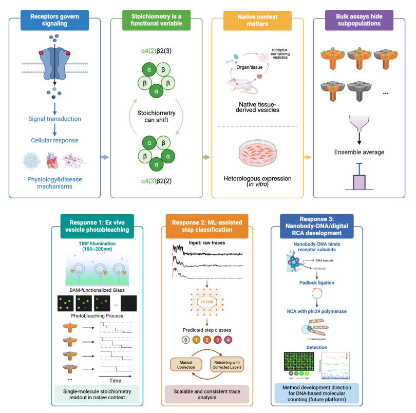
This dissertation developed quantitative strategies for measuring receptor subunit assembly at the single molecule level while preserving native tissue context. Using Ryanodine Receptor 2 (RyR2) as a tetrameric model receptor, this work established an ex vivo vesicle photobleaching platform for analyzing subunit assembly in cardiac and neuronal tissue-derived samples.
The results supported predominantly tetrameric RyR2 assembly across heart and brain regions, while also revealing a subtle cerebellum-associated deviation from the tetrameric expectation. This work demonstrated the method's capability to detect receptor subunit assembly patterns that would be difficult to resolve from bulk measurements alone. To improve the scalability of photobleaching analysis, this dissertation also developed a machine learning trace classification workflow.
One-dimensional convolutional neural network models were trained and evaluated using experimental photobleaching traces. This study showed that machine learning models can support step classification and that labeled real traces are critical for reliable performance on practical application.
To further expand the accessibility of receptor subunit counting, this dissertation explored a DNA barcode and rolling circle amplification strategy that uses nanobodies as molecular bridges between receptor subunits and amplifiable DNA probes.
This design aimed to shift the single molecule readout from detecting small stepwise fluorescence decreases during photobleaching to detecting an increasing fluorescence signal generated by DNA amplification, reducing dependence on advanced microscopy and make subunit counting more compatible with common molecular biology platforms.
Together, these studies established a workflow for receptor subunit stoichiometry analysis in tissue-derived vesicles and identify computational and molecular strategies for making single-molecule measurements more scalable and broadly applicable.