Research into starch metabolism and the fatal neurodegenerative epilepsy called Lafora disease are surprisingly linked by a family of enzymes that we recently discovered called glucan phosphatases. Plants release the energy in transitory starch via a recently identified three-step process: starch phosphorylation, degradation, and dephosphorylation. Dikinases phosphorylate the outer starch glucose units to make them water-soluble and enzyme accessible so that amylases can release maltose and glucose. Following amylase activity, the phosphate must be removed by glucan phosphatases. In the absence of glucan phosphates, plants cannot access the energy stored in starch and the starch granules grow in size while plant growth is stunted.
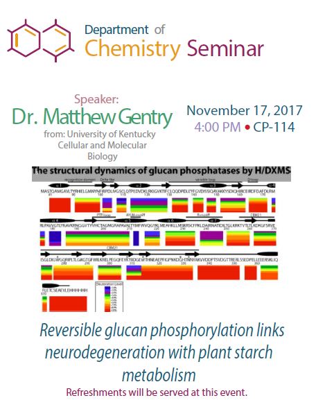
We determined the structure of the Arabidopsis glucan phosphatases Starch Excess 4 (SEX4) and Like Sex Four2 (LSF2) with phospho-glucan product bound at 1.62Å and 2.3Å, respectively. The glucose moiety closest to the SEX4 active site is positioned with the oxygen of the C6 carbon directed into the catalytic cleft and we find that SEX4 preferentially dephosphorylates C6 hydroxyls. Alternatively, the glucose moiety closest to the LSF2 active site is positioned with the C3 carbon towards the catalytic cleft and we find that LSF2 exclusively dephosphorylates C3 hydroxyls. Using structural and biochemical insights, we completely reversed SEX4 specificity, providing a method for engineering glucan phosphatase activity.
The human EPM2A gene encodes laforin and recessive mutations in EPM2A result in Lafora disease (LD). In the absence of laforin activity, glycogen transforms into a hyper-phosphorylated, water-insoluble, starch-like Lafora body (LB) that drives neuronal apoptosis, neurodegeneration, and eventual death of LD patients. The physiological function of laforin is to dephosphorylate glycogen, yet the mechanism of glycogen dephosphorylation by laforin was unknown. Additionally, LD missense mutations are dispersed throughout laforin, bringing to question the structural mechanism(s) of disease. We determined the crystal structure of human laforin at 2.4 Å bound to oligosaccharides with a phospho-glucan product at the active site. The structure reveals an integrated tertiary structure of the carbohydrate binding module and dual specificity phosphatase domains as well as an antiparallel dimer mediated by the phosphatase domain that results in a tetramodular architecture, positioning the two active sites ~31 Å from each other. We utilized the crystal structure and three solution-based, biophysical techniques along with biochemical analyses of LD patient mutations and structured guided mutations to probe this unique tertiary and quaternary structure. We define a cooperative mechanism of action for laforin as well as establish the effect of LD disease mutations, thereby providing atomic level insights that connect basic glycogen metabolism to human neurodegenerative disease.
These structures are the first of glucan phosphatases, and they provide new insights into the molecular basis of this medically-, agriculturally-, and industry-relevant enzyme family as well as their unique mechanisms of catalysis, substrate specificity, and interaction with glucans.